With the support of CIQTEK Scanning NV Microscopy (SNVM), researchers at Tsinghua University have directly visualized nanoscale spin cycloid structures in multiferroic BiFeO₃. This work, published in Advanced Functional Materials, provides the missing microscopic evidence linking crystal symmetry, magnetic structure, and anisotropic magnon transport, highlighting SNVM as a decisive tool for magnonics and low-power spintronic research.
 The study used the CIQTEK Scanning NV Probe Microscope (SNVM)
Research Background: Magnon Transport in Multiferroic Oxides
Magnon-mediated spin currents can propagate in magnetically ordered insulators with nearly zero energy dissipation, making them highly attractive for next-generation low-power spintronic devices. In multiferroic materials such as BiFeO₃, the coupling between ferroelectric and antiferromagnetic orders enables electric field control of magnons, a long-standing goal in spintronics.
Despite this promise, the microscopic origin of weakly anisotropic magnon transport in rhombohedral phase BiFeO₃, commonly referred to as R-BFO, has remained unresolved. Addressing this challenge requires direct real-space characterization of nanoscale magnetic structures, which has long been inaccessible using conventional techniques.
Technical Bottleneck: Lack of Direct Magnetic Structure Evidence
Theoretical studies have predicted that R-BFO hosts a cycloidal spin structure that plays a critical role in suppressing strong anisotropy in magnon transport. However, experimental confirmation has been elusive.
Traditional characterization techniques, such as X-ray magnetic linear dichroism, provide spatially averaged magnetic information and are unable to resolve nanoscale spin textures. As a result, the logical connection between crystal symmetry, magnetic structure, and magnon transport remained incomplete due to the absence of direct microscopic magnetic imaging.
CIQTEK SNVM Approach: Direct Nanoscale Magnetic Imaging
CIQTEK Scanning NV Microscopy (SNVM) overcomes these limitations by combining nanometer-scale spatial resolution with electron spin level magnetic field sensitivity. This enables non-invasive, quantitative imaging of local magnetic fields generated by complex spin textures inside functional materials.
In this work, the research teams led by Prof. Yi Di from the State Key Laboratory of New Ceramic Materials and Prof. Nan Tianxiang from the School of Integrated Circuits at Tsinghua University employed CIQTEK SNVM magnetic imaging to directly probe the intrinsic magnetic structure of R-BFO.
Key Findings Enabled by SNVM Magnetic Imaging
Using CIQTEK SNVM, the researchers clearly observed a uniform cycloidal spin structure within R-BFO, with a characteristic periodicity of approximately 70 nanometers. The high spatial resolution of SNVM allowed precise quantification of the cycloid wavelength and confirmed that the magnetic structure exists in a single-domain state.
By correlating SNVM nanoscale magnetic imaging with piezoresponse force microscopy, the team further demonstrated that the propagation vector k of the spin cycloid is perpendicular to the ferroelectric polarization direction P. This result provides direct experimental validation that the Dzyaloshinskii-Moriya interaction stabilizes the cycloidal spin structure in R-BFO.
These observations conclusively verify long-standing theoretical predictions and establish a complete experimental link between crystal symmetry, magnetic structure, and anisotropic magnon transport.
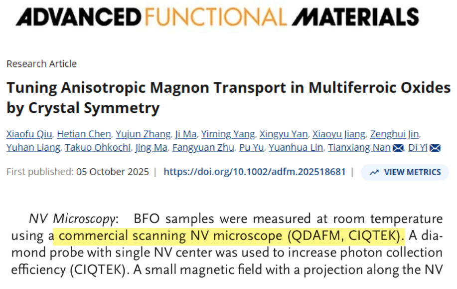
 Magnetic structures of single-domain R-BFO and O-LBFO
a) X-ray magnetic linear dichroism (XMLD) spectra of the LSMO (22 nm)/R-BFO (10 nm) sample.
b) NV imaging of the LSMO (2 nm)/R-BFO (10 nm) sample. A 2 nm-thick LSMO layer was chosen to minimize interference from its stray magnetic fields.
d) XMLD spectra of the LSMO (22 nm)/O-LBFO (10 nm) sample.
e) X-ray magnetic linear dichroism–photoemission electron microscopy (XMLD-PEEM) imaging of the LSMO (22 nm)/O-BFO (10 nm) sample.
Scientific Impact and Application Value
Published in Advanced Functional Materials under the title Tuning Anisotropic Magnon Transport in Multiferroic Oxides by Crystal Symmetry, this study significantly advances the understanding of magnon transport mechanisms in multiferroic oxides.
More importantly, it demonstrates that Scanning NV Microscopy (SNVM) is not merely a supplementary technique, but a key enabling platform for modern condensed matter physics and functional materials research. Compared with traditional spectroscopic approaches, SNVM magnetic imaging uniquely provides direct, real-space access to complex magnetic textures at the nanoscale.
Looking forward, CIQTEK SNVM is expected to play an increasingly important role in multiferroic materials, antiferromagnetic devices, and magnon-based information processing, accelerating the development of energy-efficient spintronic technologies.
Experience CIQTEK SNVM for Nanoscale Magnetic Imaging
The CIQTEK Scanning NV Microscope (SNVM) is a state-of-the-art nanoscale magnetic field imaging system designed for advanced materials research. It supports temperatures from 1.8 to 300 K, vector magnetic fields up to 9 T out of plane and 1 T in plane, spatial resolution down to 10 nm, and magnetic sensitivity reaching 2 μT per square root Hertz.
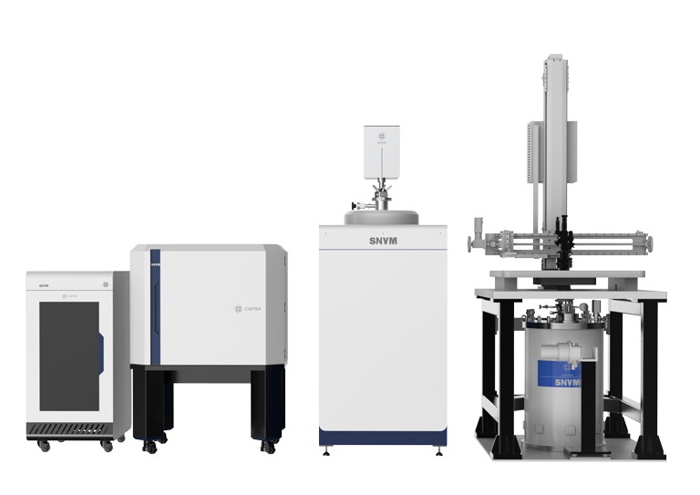 CIQTEK Scanning NV Microscope (SNVM) has two versions: the ambient version and the cryogenic version
